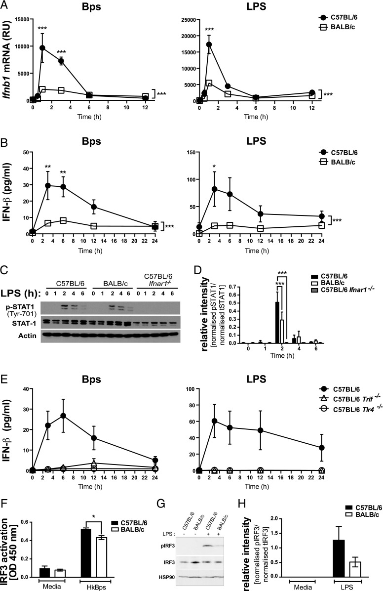
FIGURE 3.
TLR4-dependent IFN-β production and STAT1 and IRF3 activation are higher in C57BL/6 compared with BALB/c macrophages. BMDMs were stimulated with B. pseudomallei or LPS for the indicated times. (A) Ifnb1 mRNA expression was determined by qRT-PCR and normalized to Hprt1 mRNA expression. (B) IFN-β production was quantified by ELISA. (C) Whole-protein extracts were generated and analyzed by Western blot for total and phosphorylated STAT1, and actin loading control. (D) Relative intensity of two independent experiments shown for data represented in (C). (E) IFN-β production was quantified by ELISA. (F) C57BL/6 and BALB/c macrophages were stimulated with B. pseudomallei for 2 h, and nuclear extracts were analyzed for active IRF3 by ELISA. (G) Whole-protein extracts were generated and analyzed by Western blot for total and phosphorylated IRF3 and heat shock protein 90 loading control. (H) Relative intensity of three independent experiments shown from data in (G). Graphs show means ± SEM of two to four (E) or at least three independent experiments (A and B). *p < 0.05, **p < 0.01, ***p < 0.001 as determined by two-way ANOVA (Bonferroni multiple comparison test).

