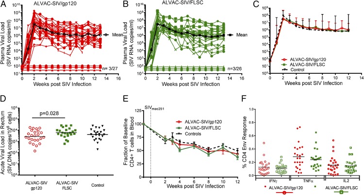
FIGURE 5.
Lack of virus control in ALVAC-SIV–primed gp120 or FLSC boosted infected macaques. (A) SIV plasma virus in ALVAC-SIV/gp120 animals over the course of the study; the geometric mean of all SIV-infected animals is shown in black. Three of 27 animals remained SIV− (SIV RNA < 50 copies/ml) over the 13 wk of follow-up. (B) SIV plasma virus in ALVAC-SIV/FLSC animals over the course of the study; the geometric mean of all SIV-infected animals is shown in black. Three of 26 vaccinated animals remained SIV− (SIV RNA < 50 copies/ml) over the 13 wk of follow-up. (C) Comparison of the geometric mean of plasma virus in SIV-infected animals from the gp120 group (red), FLSC group (green), and controls (black). No difference in peak or set point plasma virus is observed between vaccinated animals and controls. (D) Virus burden in the rectal mucosa measured as SIV DNA/106 mononuclear cells. Rectal biopsies were obtained 3 wk post SIV infection, and the ALVAC-SIV/gp120 animals are shown in red open circles, the ALVAC-SIV/FLSC animals in green squares, and the controls in black triangles. A significantly lower level of SIV DNA was observed in the gp120 group compared with the FLSC group (p = 0.028), but when both vaccine groups and controls are compared the differences are not significant (p = 0.074). (E) Percentage of baseline CD4 T cells in the blood over the course of the study. A similar progressive loss of CD4 T cells is observed post SIV infection over the 12 wk of follow-up in vaccinated animals (red lines, gp120 group; green lines, FLSC group) and controls (black line). (F) Envelope (Env)-specific CD4+ T cell responses measured in PBMCs 1 wk before SIV challenge. Shown is the frequency of IFN-γ+, TNF-α+, or IL2+ T cells after stimulation with overlapping SIVmac251-m766 Env peptides. ALVAC-SIV/gp120–vaccinated animals are shown by circles, whereas ALVAC-SIV/FLSC animals are shown by squares. A similar frequency of Env-specific CD4 T cells is observed in both groups after background subtraction of unstimulated cells. The data for the ALVAC-SIV/gp120 group (A and C–F) have been reported previously (24).

