
Figure 1.
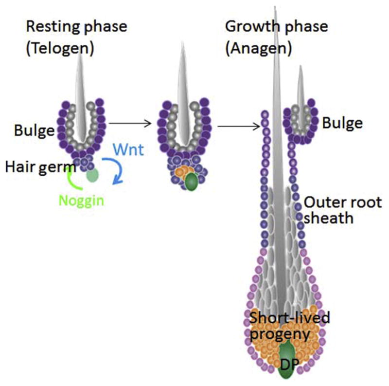
Schematic of the resting and growth phases of the hair follicle. The stem cells (purple) reside in the outer layer of the bulge niche, which anchors the hair from the previous hair cycle. The bulge cells at the base are referred to as the hair germ, which during telogen, is abutted next to the dermal papilla (DP), the mesenchymal stimulus of the hair follicle. Following a buildup of BMP inhibitors (Noggin) in the DP and WNTs in the hair germ, the stem cells at the base are activated and generate the short-lived progeny (orange), which ultimately will differentiate into the hair and its channel. This figure is patterned after Figure S5 in Hsu, Li, and Fuchs (2014b). Elsevier Press.