
Figure 2.
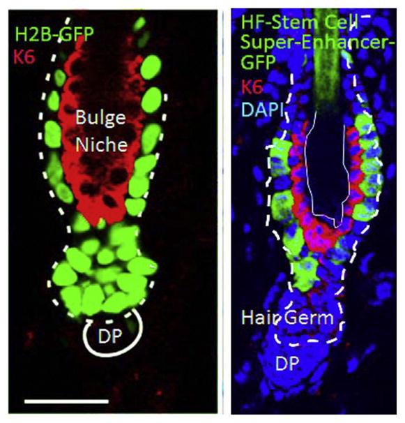
Immunofluorescence images showing a resting phase hair follicle, replete with bulge stem cells and keratin 6+ inner bulge nonstem cells. At left, bulge and hair germ cells are label-retaining and marked by H2B-GFP used in a pulse-chase labeling experiment. At right, bulge is labeled with GFP driven by a HF stem cell-specific super-enhancer epicenter element. Left panel is Figure 5 Frame C of Hsu et al. (2011). Elsevier Press. Right panel is Figure 2e of Adam et al. (2015). Nature Press.