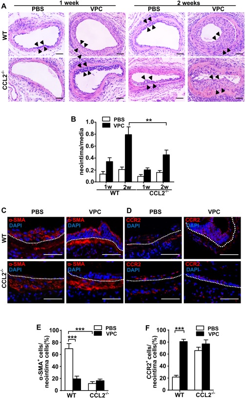
Figure 6.

Lack of CCL2 reduces neointima formation and inhibits stem cell antigen‐1 cells migration and differentiation into smooth muscle cell (SMCs). (A): Animals were euthanized at indicated time points after injury, and the femoral arteries were fixed in 4% phosphate‐buffered (pH = 7.2) formaldehyde, embedded in paraffin, sectioned in 5 µm, and stained with hematoxylin–eosin. Scale bars, 50 µm. (B): The ratio of neointima (the area between arrows) to media was quantified as shown in the graph. (C, D): Vessel sections were also prepared for immunofluorescent α‐SMA and CCR2 staining 2 weeks postwire injury (white dotted line indicates internal elastin, and the above is neointima area). Scale bars, 50 µm. (E, F): Quantification of the percentage of positively stained cells within the neointima was shown in graphs as mean ± SEM of n = 8 mice/group. **p < .01, ***p < .001. Abbreviations: PBS, phosphate buffered saline; VPC, vascular progenitor cell; WT, wild type.
