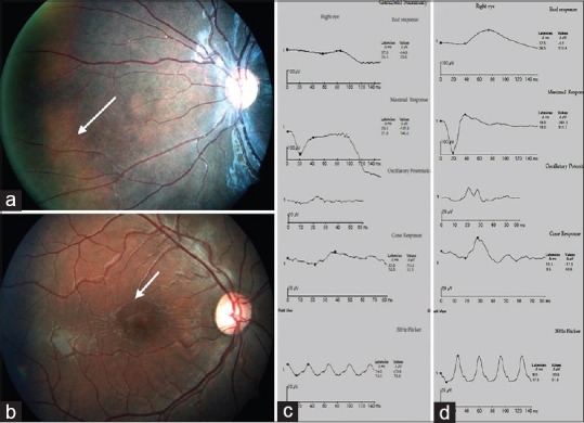
Figure 2.

(a) Fundus picture depicting tapetal reflex. (b) Foveal schisis seen on fundus photography. (c) Full field electroretinogram showing reduced waveforms on all recordings. Combined response shows a negative waveform in a case of foveal + peripheral schisis. (d) Full field electroretinogram in a case of foveal schisis shows better waveforms compared to the peripheral schisis scenario
