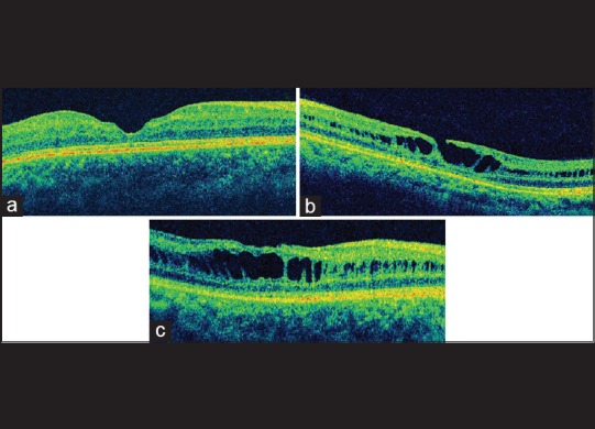
Figure 5.

(a) Raster optical coherence tomography scan shows a foveal thinning with intact photoreceptor layer. (b) Lamellar macular hole with inner segment/outer segment defects of the photoreceptors. (c) Optical coherence tomography image showing an abnormal and flat foveal contour along with retinal pigment epithelium alterations
