
Figure 1.
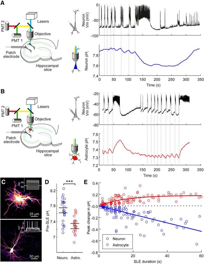
Astrocytes experience rapid intracellular alkalinization during epileptiform activity. A, Schematic (left) showing the experimental setup in which a hippocampal pyramidal neuron expressing a genetically encoded pH reporter was imaged, while a simultaneous patch-clamp recording was performed from a neighboring neuron. Dynamic intracellular pH measurements from a representative CA3 pyramidal neuron expressing E2GFP (right, blue trace) and current-clamp recording from a neighboring pyramidal neuron (black trace; somata <200 μm apart), which provided a readout of the epileptiform activity in Mg2+-free ACSF. Acidic intracellular pH shifts were closely associated with SLEs (onset indicated by vertical dashed lines). B, Schematic (left) showing a similar setup as in A for the measurement of pH dynamics in hippocampal astrocytes during SLEs. Intracellular pH measurements made from a representative astrocyte expressing E2GFP (right, red trace, same astrocyte as in C) and current-clamp recording from a neighboring pyramidal neuron (black trace), which provided a readout of the epileptiform activity in Mg2+-free ACSF. Individual SLEs of different durations are closely associated with rapid intracellular alkaline transients in the astrocyte. C, Confocal image (top) of an astrocyte expressing E2GFP. After performing pH imaging (shown in B), this astrocyte was targeted for whole-cell patch-clamp recording and confirmed to exhibit a low membrane resistance (Rm = 20.5 MΩ) and lack voltage-gated conductances in response to current injection (inset). Confocal image (bottom) of a neuron expressing E2GFP, which exhibited action potentials in response to current injection (inset). D, Population data demonstrating a significant difference in pre-SLE intracellular pH between hippocampal neurons and astrocytes. ***p < 0.0001, t test. E, Population data showing the peak shift in intracellular pH as a function of the duration of the epileptiform activity (data from 23 neurons, blue, and 24 astrocytes, red). For neurons, the peak change in pH was negatively correlated with the duration of the SLE (r = −0.6719, p < 0.0001, Pearson correlation). For astrocytes, the peak pH shift was positively correlated with the length of the SLE (r = 0.5766, p < 0.0001, Pearson correlation).