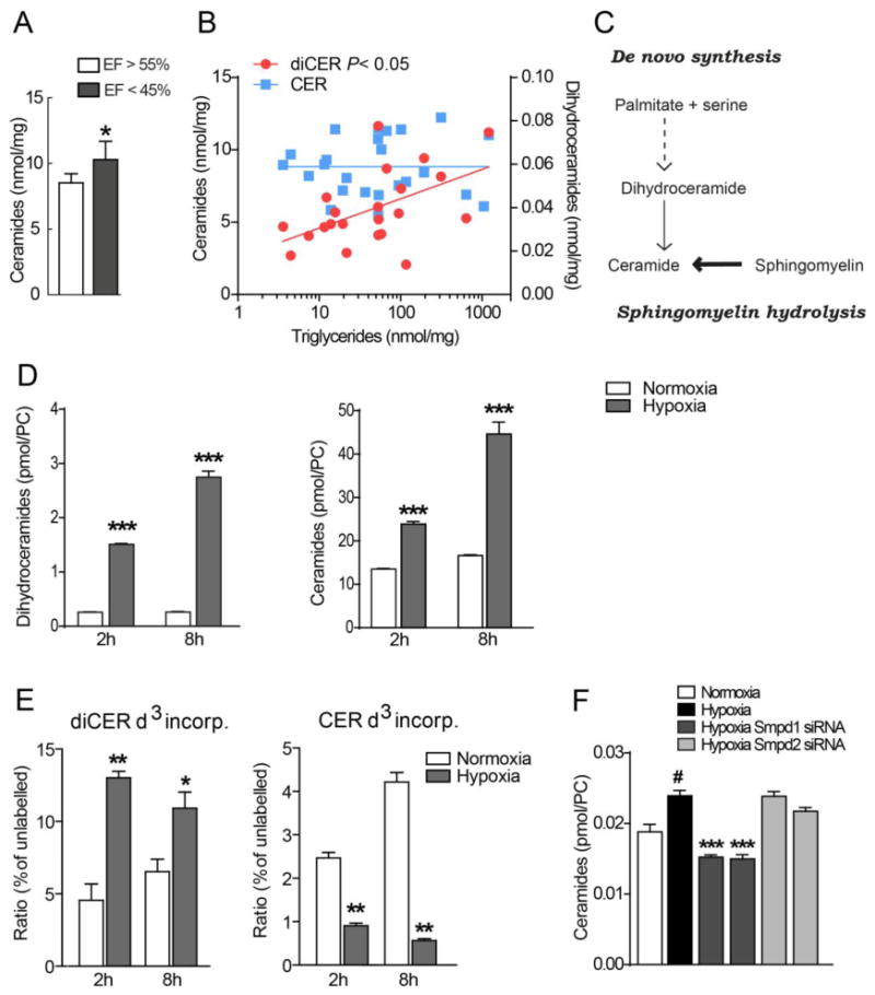
Figure 1. Ceramides correlate with heart function and hypoxia-induced ceramide accumulation in cardiomyocytes is mediated by acid sphingomyelinase.

(A) Ceramide concentrations in heart biopsies from patients with chronic cardiac ischemia and ejection fraction (EF) >55% or <45% (n=30). * P<0.05, vs EF>55%. (B) Correlations between ceramides and dihydroceramides with triglycerides in heart biopsies from patients with chronic cardiac ischemia (n=30). (C) Ceramide synthesis pathway. (D) Hypoxia-induced ceramide accumulation in HL-1 cardiomyocytes. HL-1 cells were incubated in normoxia or hypoxia for 2 h or 8 h and dihydroceramides and ceramides were measured with HPLC and mass spectrometry. (n = 3). Data are shown as mean ± SEM, ***P < 0.001 vs normoxia. (E) Incorporation of deuterium-labelled serine into dihydroceramides and ceramides in HL-1 cardiomyocytes following incubation in normoxia or hypoxia for 2 h or 8 h (n=3) ***P < 0.001 vs normoxia. (F) Ceramide levels in HL-1 cardiomyocytes transfected with control siRNA, Smpd1 siRNA (ID #1 and #2) and Smpd2 siRNA (ID #1 and #2), incubated in normoxia or hypoxia for 24 h (n = 3–6). Data are mean ± SEM.
