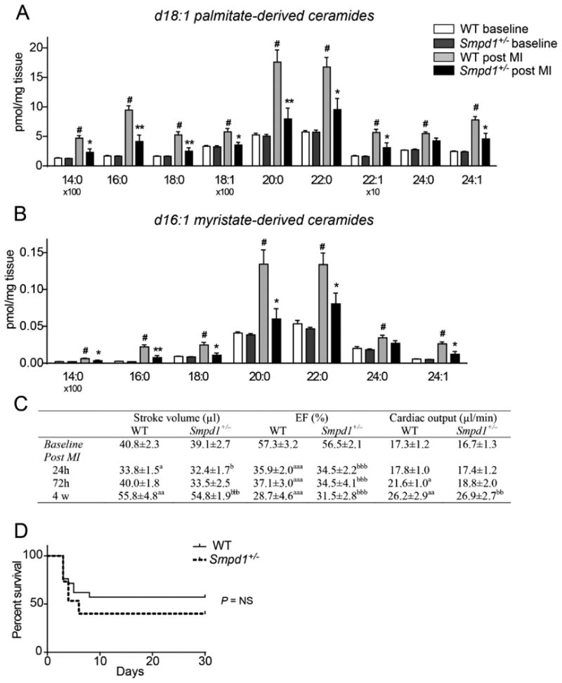
Figure 2. Inactivation of acid sphingomyelinase (Smpd1) reduces ischemia-induced cardiac ceramide accumulation but does not affect heart function.

(A–B) Concentrations of d18:1 palmitate-derived (A) and d16:1 myristate-derived (B) ceramide species in WT and Smpd1+/– mice at baseline and 24 h after an induced myocardial infarction (MI) (n=5–6). Data are mean ± SEM. #P < 0.01 vs WT baseline *P < 0.05, **P < 0.01 vs WT post MI. (C) Echocardiographic analysis of WT and Smpd1+/– mice at baseline and after an induced MI (n=9 at baseline; n=15–20 at 24 h; n=7–9 at 72 h; n=6–7 at 4 weeks). aP < 0.05, aaP < 0.01 and aaaP < 0.001 vs WT baseline; bP < 0.05, bbP < 0.01 and bbbP < 0.001 vs Smpd1+/– baseline. (D) Kaplan-Meier curve showing survival of WT and Smpd1+/– mice after an induced MI (n=15–21). Data are analyzed using the log-rank test and P = 0.33.
