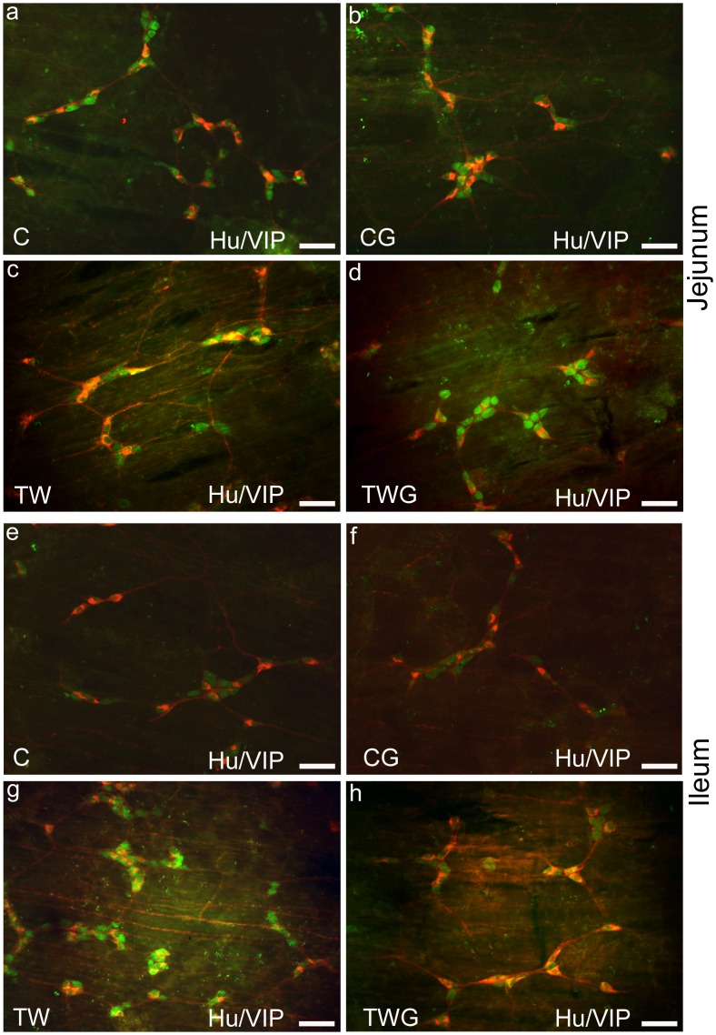
Fig 2. Representative photomicrographs of Hu/VIP submucous neurons.
Double immunostaining of HuC/D and VIP submucous neurons of jejunum (a-d) and ileum (e-f) from experimental groups: control (C); control supplemented with 2% L-glutamine (CG); Walker-256 tumor (TW); and Walker-256 tumor supplemented with 2% L-glutamine (TWG). These images are composites of merged images taken separately from red (VIP) and green (HuC/D) fluorescent channels. Scale Bar 50 μm.

