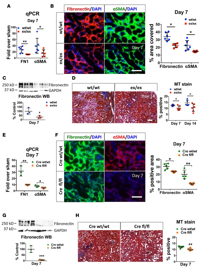
Figure 3. ADAM17ex/ex and ADAM17 PTC-KO mice are protected from UUO-induced fibrosis.
Mice were subjected to UUO, and their kidneys were examined 7 or 14 days after ureteral ligation, as noted. (A) qPCR analysis of fibronectin or αSMA mRNA expression levels in whole kidneys of ex/ex or WT/WT mice at day 7, expressed as fold over respective sham-injured mice (n = 6). (B) The induction of fibronectin and αSMA in the injured kidney cortex of UUO-subjected WT/WT or ex/ex mice was examined by immunostaining (left: representative images; right: quantification; n = 6; scale bar: 50 μm). (C) Fibronectin protein expression at day 7 after ligation was examined by Western blot (top: sample blots; bottom: quantification; GAPDH was used as loading control; n = 3). (D) Masson’s trichrome staining in kidney cortex at day 7 or day 14 after ligation (left: representative images day 14; right: quantification; n = 5–6; scale bar: 100 μm). (E) qPCR analysis of fibronectin or αSMA mRNA expression levels in whole kidneys of UUO-subjected control (Cre WT/WT) or ADAM17 PTC-KO (Cre fl/fl) at day 7, expressed as fold over respective sham-injured mice (n = 3). (F) The induction of fibronectin and αSMA in the injured kidney cortex of UUO-subjected control (Cre WT/WT) or ADAM17 PTC-KO (Cre fl/fl) mice was examined at day 7 by immunostaining (left: representative images, right: quantification; n = 5; scale bar: 50 μm). (G) Fibronectin protein expression at day 7 after ligation was examined by Western blot (top: sample blots; bottom: quantification; GAPDH was used as loading control; n = 3). (H) Masson’s trichrome staining in kidney cortex at day 7 after ligation in control (Cre WT/WT) or ADAM17 PTC-KO (Cre fl/fl) mice (left: representative images; right: quantification; n = 6; scale bar: 100 μm). *P < 0.05; **P < 0.01; ***P < 0.001 as determined by an unpaired 2-tailed Student’s t test. ADAM17; a disintegrin and metalloprotease 17; FN1, fibronectin; MT, Masson’s trichrome; PTC-KO, proximal tubule cell KO; αSMA, alpha smooth muscle actin; UUO, unilateral ureteral obstruction; WB, Western blot.

