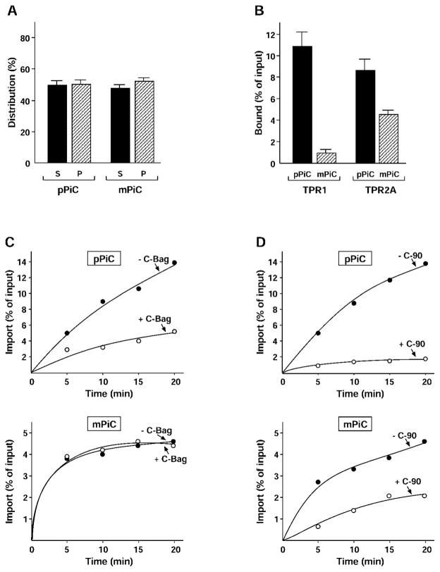
Figure 2. The PiC presequence determines Hsc70 interactions.
(A) pPiC and mPiC were radiolabelled by cell-free translation in reticulocyte lysate, reactions were terminated and separated by centrifugation. The supernatants (S) and pellets (P) were analysed by SDS/PAGE, fluorography and densitometry, and the distribution as a percentage of total protein is shown. Results are means ± S.D. (n ≥3). (B) pPiC and mPiC were radiolabelled and incubated with purified His-tagged Hop TPR1 and TPR2A fragments. The fragments were recovered with nickel–Sepharose and the co-precipitated protein was analysed. The amount of binding is shown as a percentage of input translation. Results are means ± S.D. (n ≥3). (C) pPiC and mPiC were radiolabelled and incubated with isolated rat liver mitochondria for the indicated times to allow import. Mitochondria were re-isolated and treated with proteinase K, and the protease-resistant imported material was analysed. Reactions were conducted in the absence (− C-Bag, ●) or presence of C-Bag fragment (+ C-Bag, ○). Import is shown as a percentage of input translation. (D) The import of pPiC and mPiC was assayed as stated for (C), except that reactions contained either no addition (− C-90, ●) or purified C-90 fragment (+ C-90, ○).

