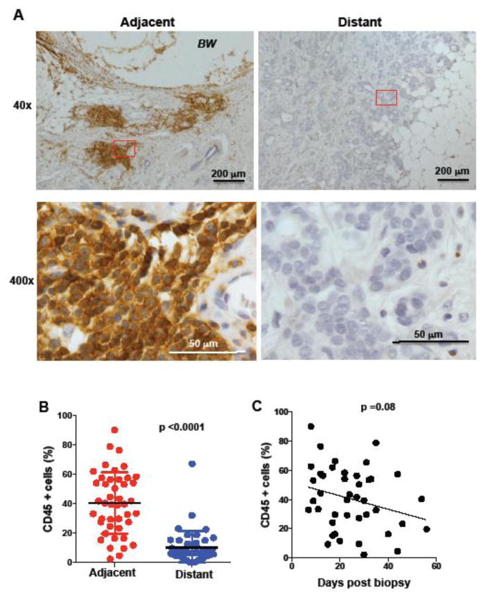
Figure 1. Accumulation of inflammatory cells at the region adjacent to the biopsy wound.
(A) IHC of human breast tissue for CD45 marker in regions of the tumor adjacent to the biopsy wound (adjacent) and regions far away from the biopsy wound (distant). The images 40x magnification (upper panels) and 400x magnification (lower panels) of the 40x-red labeled areas are shown. Scale bars are shown. BW, biopsy wound. (B) Percentage of CD45 positive cells (relative to all nucleated cells in the analyzed fields) in breast cancer samples (n= 44) in regions adjacent and distant to the biopsy wound, p<0.0001 using a paired student’s t test analysis. (C) Percentage of CD45 positive cells in in the region adjacent to the biopsy as a function of time (days) between the core needle biopsy procedure and surgical excision of the breast tumor tissue (n=44), p=0.08 as determined by regression analysis.

