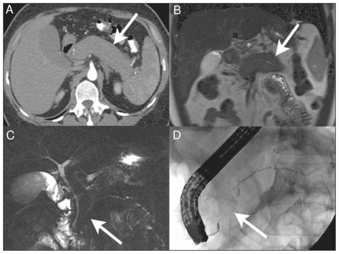
Figure 3:
Diffuse autoimmune pancreatitis.68 (A) Axial computed tomography (CT) image in the pancreatic parenchymal phase of the typical enlarged and poorly enhanced pancreas in a patient with diffuse autoimmune pancreatitis68 (arrow). Note the lack of inflammatory change around the organ, which differentiates the disease from acute pancreatitis and necrosis. (B) View of the pancreas using coronal T2-weighted magnetic resonance imaging (MRI) that shows low signal intensity in the pancreas (arrow) because of the diffuse fibrosis in the gland. (C) Coronal magnetic resonance cholangiopancreatography image showing a diffusely irregular pancreatic duct with stenosis distally in the pancreatic head (arrow). (D) Endoscopic retrograde cholangiopancreatography image that confirms the MRI findings, including ductal stenosis (arrow). Images reproduced from reference 68 under Creative Commons licence 2.0 (http://creativecommons.org/licenses/by/2.0/legalcode).

