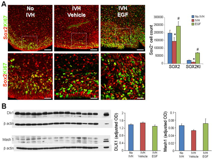
Fig. 8. rhEGF treatment induces proliferation of Sox2+ cells and does not affect Dlx1, Mash1 proteins.
A) Representative immunofluorescence of cryosections from pups (D3) labeled with Sox2 and Ki67 antibodies. Lower panels are high magnification images of the boxed area in the upper panel. Note reduced density of total and proliferating (arrowhead) Sox2 in pups with IVH and increase in their density on rhEGF treatment. Data are mean ± s.e.m. Scale Bar, 50 μm (upper panel), 20μm (lower panel). B) A typical Western blot analyses of Dlx1, and Mash1 performed on forebrain homogenates of pups at D3. Adult rat brain was positive-control. Graph shows mean ± s.e.m. (n = 5 each). Values were normalized to β-actin. rhEGF treatment did not affect Dlx1 and Mash1 levels. *P < 0.05 for no IVH vs. IVH. #P < 0.05, for vehicle treated vs. rhEGF treated pups with IVH.

