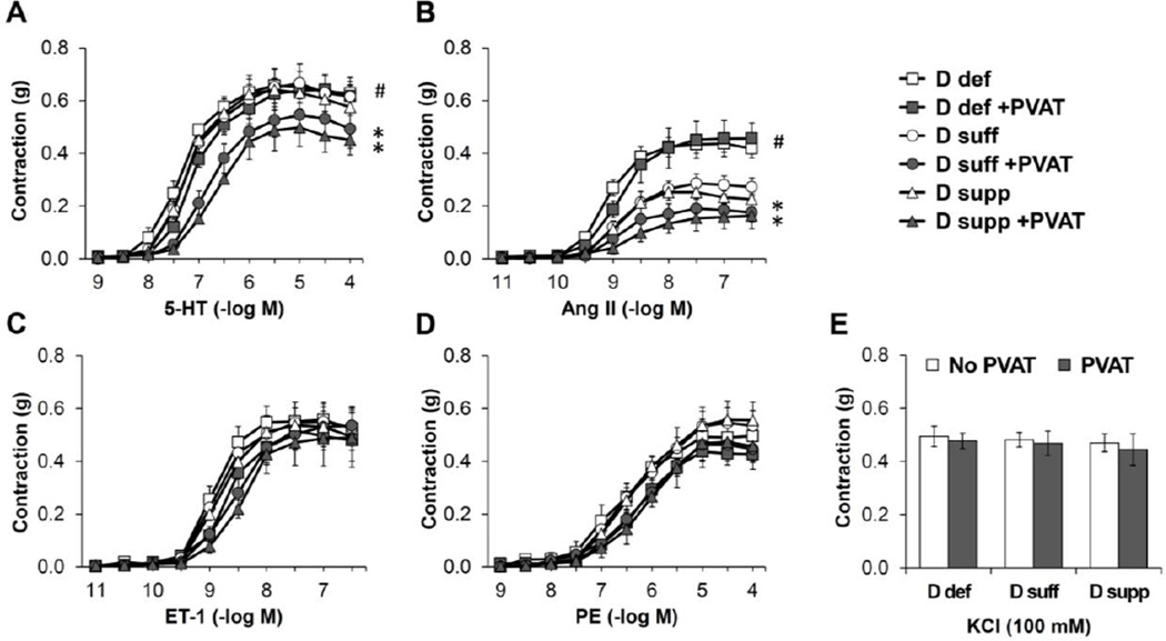
Figure 2. Vitamin D deficiency eliminates anti-contractile effects of PVAT in mesenteric resistance arteries.
Isometric tension was measured on second order mesenteric arteries from mice maintained on vitamin D-deficient diet (D def; squares), vitamin D-sufficient diet (D suff; circles) or vitamin D-supplemented diet (D supp; triangles). The mesenteric artery rings were either dissected free of PVAT (open symbols) or had the PVAT remaining intact (filled symbols). Dose-dependent contraction was assessed in response to 5-HT (A), angiotensin II (Ang II) (B), endothelin-1 (ET-1) (C), PE (D) or KCl (E). Data represent means ± SE (n=5 mice). *p<0.05 PVAT vs. no PVAT. #p<0.05 D def or D supp vs. D suff.

