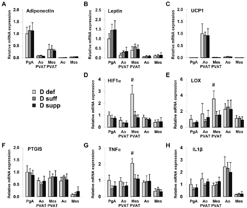
Figure 4. Vitamin D deficiency upregulates mediators of hypoxia and inflammation in mesenteric PVAT.
Gene expression analysis was performed on samples of perigenital adipose tissue (PgA), aortic PVAT (Ao PVAT), mesenteric PVAT (Mes PVAT), aortic tissue (Ao) and mesenteric artery tissue (Mes) by quantitative real-time PCR. Relative expression of mRNA transcripts was determined after normalization to GAPDH. Data for adiponectin (A), leptin (B), HIF-1α (D), LOX (E), PTGIS (F), TNFα (G) and IL1β (H) are fold-change of vitamin D-deficient perigenital adipose tissue. UCP1 data (C) are fold change of vitamin D-deficient aortic PVAT. Data represent means ± SE (n=6 mice). #p<0.05 D def or D supp vs. D suff.

