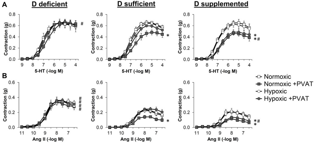
Figure 6. Hypoxic incubation inhibits anti-contractile effects of PVAT in normal mesenteric arteries.
Isometric tension was measured on first order mesenteric arteries from mice maintained on the three vitamin D diets. The mesenteric artery rings were either dissected free of PVAT (open symbols) or had the PVAT remaining intact (filled symbols) and incubated for 24 hours under normoxic (squares) or hypoxic conditions (circles). Dose-dependent contraction was assessed in response to 5-HT (A) and Ang II (B) (see also Figure S2). Data represent means ± SE (n=4–5 mice). *p<0.05 PVAT vs. no PVAT. #p<0.05 D def or D supp vs. D suff.

