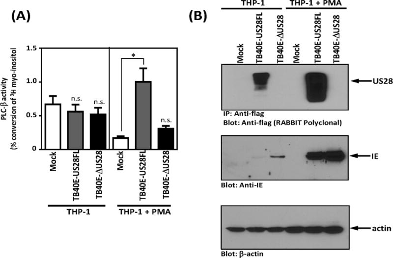
Figure 2. US28 constitutively induces PLC-β signaling in HCMV infected THP-1 cells.

(A) THP-1 and PMA differentiated THP-1 cells were mock infected or infected with TB40E-US28FL or TB40E-ΔUS28 viruses. At day 4 post-infection, cells were harvested and PLC-β activity was assessed. (B) THP-1 and PMA-differentiated THP-1 cells were mock infected or infected with the indicated TB40E viruses. US28 protein expression was analyzed by immunoprecipitation and western blot at day 4 post-infection. Data are representative of 3–4 independent experiments. Statistical significance was determined by performing unpaired two-tailed Student’s t tests with GraphPad Prism® software. **: p<0.01, *: p<0.05.
