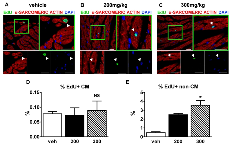
Figure 5. 200mg/kg ISO does not increase the % of EdU+ myocyte nuclei.
A-C, Confocal micrographs of fixed hearts at 1 week post-injection: veh (A); 200mg/kg ISO (B), and 300mg/kg ISO (C) that were immunostained against α-sarcomeric actin(red), 5-ethynyl-2′-deoxyuridine (EdU; green), and nuclei labeled with 4’,6-diamidino-2-phenylindole (DAPI, blue). White arrowheads=myocyte nuclei. Scale bars=25μm. D and E, Percentage of EdU+ myocyte (D) (n=9, 3 each group) or non-myocyte (E) (n=9, 3 each group) nuclei 4 weeks post-injection. Veh=vehicle; 200=200mg/kg ISO, 300=300mg/kg ISO. *p<0.05 vs. veh, NS=not significant (p>0.05). D and E, Data are shown as mean ± SEM. See also Online Figure IV.

