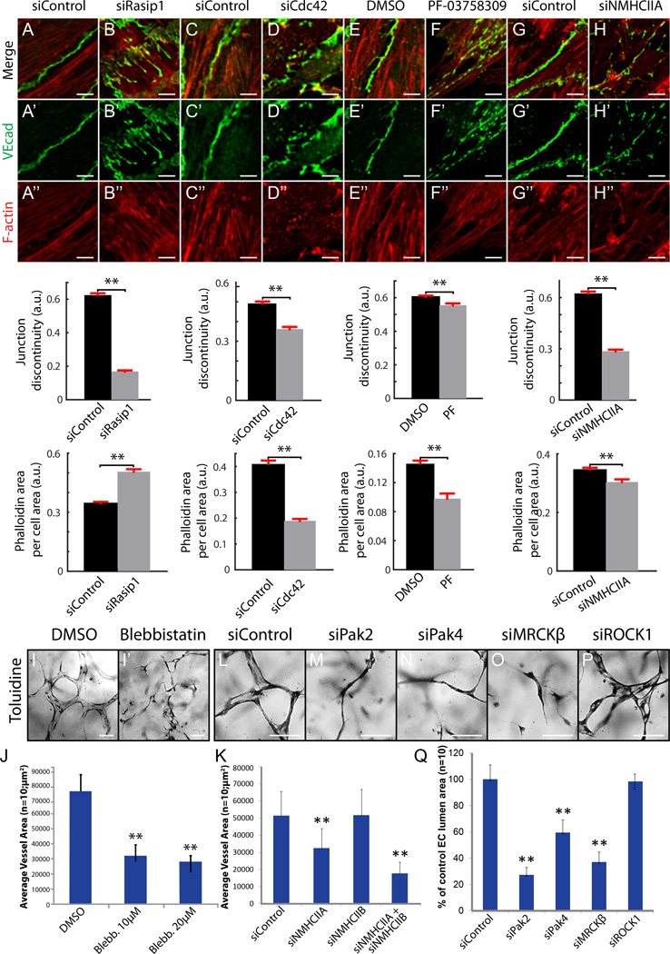
Figure 6. Cdc42, Pak4 and NMII pathway downstream of Rasip1 supports EC-EC junctions and vascular lumenogenesis.
(A–H’’) Staining and quantification of VEcad continuity and F-actin area at MS1 EC cell-cell junctions after siRNA reduction of Rasip1, Cdc42, or NMHCIIA, or pharmacological inhibition of Pak4 (n=3 control and treated, 15 FOV). **P<0.01. (I–J) Inhibition of NMII via 10µM or 20µM blebbistatin treatment prevents EC lumen formation in 3D collagen matrices (quantified in J). **P<0.01. (K) Graph showing that reduction of NMHCIIA or NMHCIIA and NMHCIIB combined, but not NMHCIIB alone, prevents EC lumen formation in 3D collagen matrices. **P<0.01. (L–XQ) siRNA reduction of Cdc42 effectors Pak2, Pak4, or MRCKβ but not RhoA effector ROCK prevents EC lumen formation in 3D collagen matrices (quantified in Q). **P<0.01. Scale bars: A–N’’ 5µm, Q–X 100µm.

