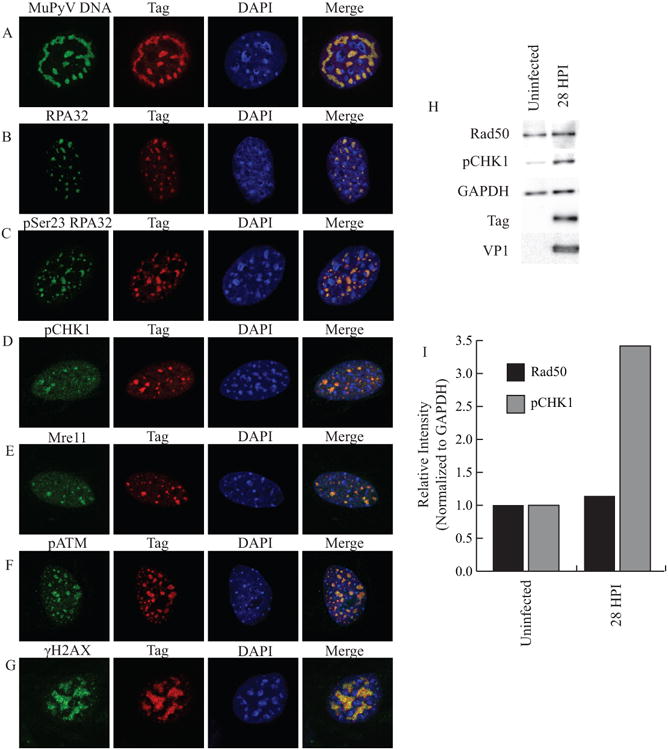
Figure 1. MuPyV activates and reorganizes host DDR proteins in the nucleus.

MEFs were infected with WT MuPyV (NG59RA), and fixed, permeabilized, and immunostained at 28 hrs post infection (HPI). Single z-plane confocal immunofluorescence images show cells stained for Tag and A) MuPyV DNA (FISH) B) RPA32 C) pSer23 RPA32 D) pSer345 CHK1 E) Mre11 F) pATM or G) γH2AX. H) Total proteins were isolated at 28 HPI and analyzed by immunoblot for pSer317 CHK1 and Rad50 protein levels. I) Quantification of integrated density of pSer317 CHK1 and Rad50 levels in control and infected MEFs normalized to GAPDH loading control. All samples were normalized to the value of the uninfected control cells.
