Fig. 1.
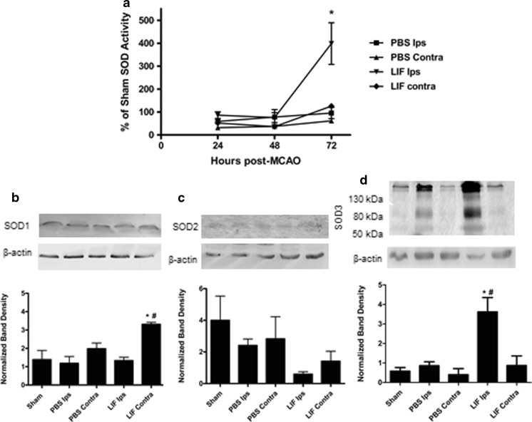
LIF increases SOD activity and SOD3 Expression. a Total SOD activity was measured in brain lysates from PBS- and LIF-treated rats euthanized 24, 48, or 72 h post-MCAO. LIF ipsilateral samples had significantly higher SOD activity compared to PBS-treated ipsilateral samples at 72 h post-MCAO (*P < 0.05). Mean activities were normalized to SOD activity in brains from sham rats. n ≥ 5 samples per group. b At 72 h after MCAO, SOD1 expression was significantly increased in contralateral tissue of LIF-treated rats, compared to ipsilateral tissue from PBS-treated rats (*P < 0.01) and ipsilateral tissue from the same treatment group (#P < 0.001). SOD1 bands were observed at approximately 18 kDa. c There was a trend towards decreased SOD2 expression in the ipsilateral samples from the LIF group; however, this decrease was not significant. SOD2 bands were observed at approximately 25 kDa. d Ipsilateral samples from LIF-treated rats showed a corresponding increase in SOD3 expression compared to PBS (*P < 0.05) and sham (#P < 0.01) ipsilateral samples. Three bands corresponding to SOD3 were observed in all samples at approximately 50, 80, and 130 kDa. n = 3 samples per group. Ips ipsilateral, Contra contralateral

