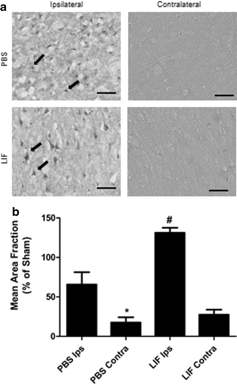
Fig. 2.
LIF upregulates SOD3 in the ipsilateral cortex. a Representative images of cortical tissue from PBS rats show higher levels of SOD3 staining compared to contralateral tissue from the same group. However, these cells appeared dysmorphic and unhealthy. LIF treatment increased SOD3 expression in the ipsilateral cerebral cortex while visibly reducing damage to SOD3-positive cells and surrounding tissue. b After normalizing to sham SOD3 levels, immunohistochemical quantification revealed significantly higher levels of staining in ipsilateral tissue compared to contralateral tissue in PBS-treated rats (*P < 0.05). LIF treatment further raised SOD3 in the ipsilateral tissue compared to PBS (#P < 0.01). n = 5 per treatment group. Scale bars = 50 μm. Ips ipsilateral, Contra contralateral

