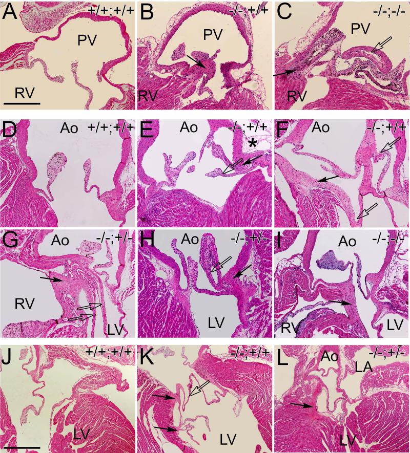
Fig. 4. Pulmonary, aortic and mitral valves in the Fmod−/−;Lum+/+, Fmod−/−;Lum+/− and Fmod−/−;Lum−/− adult hearts exhibited abnormal cusp attachment and ectopic tissue.
Hematoxylin and eosin stained sections from the pulmonary valve (A-C) aortic valve (D-I) and mitral valve (J-L) of Fmod+/+;Lum+/+ (A, D, J), Fmod−/−;Lum+/+ (B, E, F, K), Fmod−/−;Lum+/− (G, H, L) and Fmod−/−;Lum−/− (C, I) hearts from 1.5 month to 5 months old. PV- pulmonary valve; RV- right ventricle; Ao- aorta, LV- left ventricle; LA- left atrium. Solid arrows- ectopic tissue; open arrows- extra cusp/leaflet-like extensions. Scale bars: A= 280μm applies to B-I; J= 700μm applies to K and L. Movies have been added to demonstrate the three-dimensional changes that correspond to the aortic malformation shown in the Fmod−/−;Lum+/+ heart in F and the Fmod−/−;Lum+/− heart shown in panel K.

