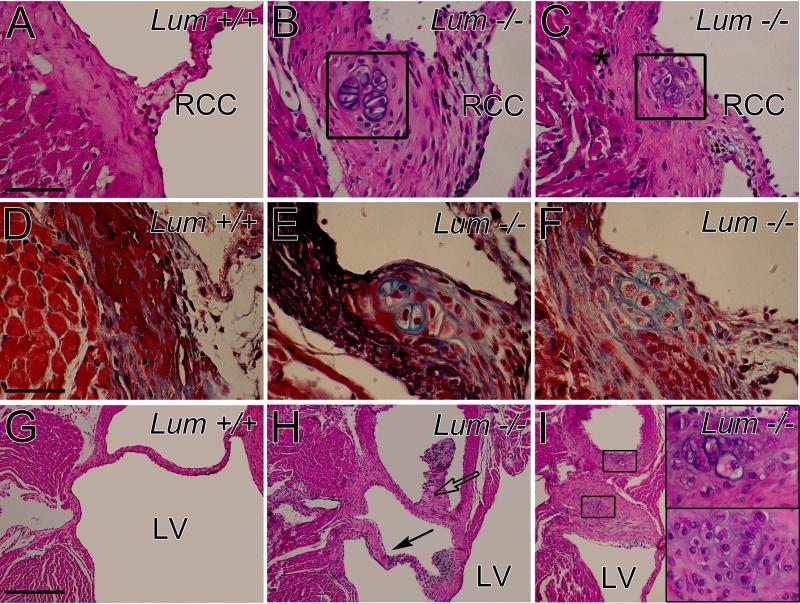
Fig. 5. Mice deficient in lumican showed cartilage-like nodules in the annulus and adjacent to aortic valve cusps.
Hematoxylin and eosin stained sections of the anchor region of the aortic valve right coronary cusp (RCC) in Lum+/+ (A) and Lum−/− (B, C) adult hearts. Movat's pentachrome stained sections (D-F) highlight cartilage-like nodules (green) in the Lum−/− (E, F) hearts. Hematoxylin and eosin stained sections of the aortic valve non coronary cusp (NCC; G, H); boxes (I) show magnified lacunae (cartilage-like regions). Solid arrow- abnormal valve extension; open arrow- extra tissue of the NCC; Ao- aorta; LV- left ventricle. Scale bars: A= 70 μm applies to B, C; D= 45μm applies to E and F, insets in I; G= 280μm applies to H and I.

