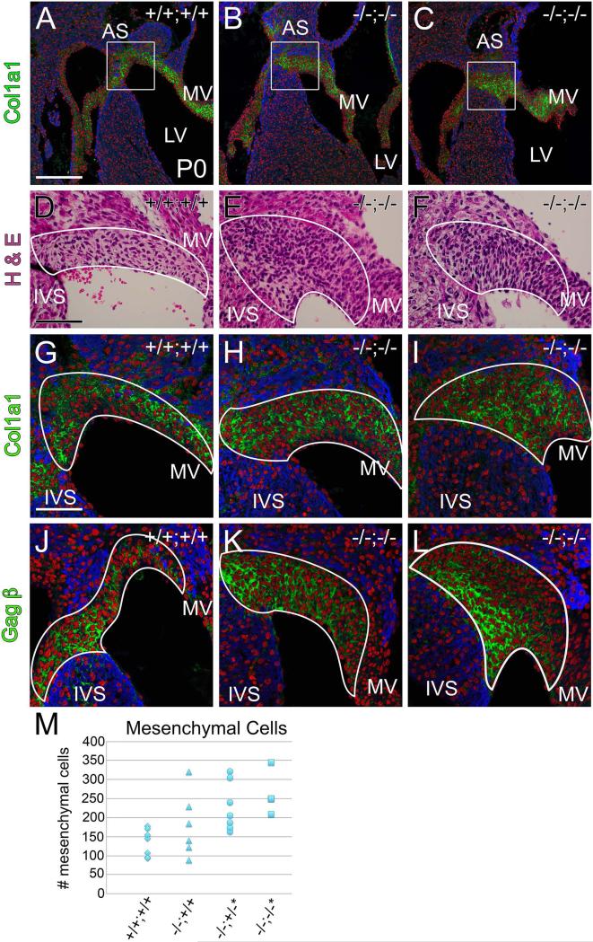
Fig. 8. Mesenchymal cell number was increased at the juncture of the mitral valve leaflet with the interventricular septum at postnatal day 0 in the Fmod−/−;Lum+/− and Fmod−/−;Lum−/− hearts.
Histological sections from Fmod+/+;Lum+/+ (A, D, G, J,) and Fmod−/−;Lum−/− (B, C, E, F, H, I, K, L) hearts are shown. Immunohistochemistry of Col1a1 in P0 hearts (green, AC; G-I) is depicted; boxes in A-C are magnified in G-I. Hematoxylin and eosin stained sections at the juncture of the interventricular septum (IVS) and mitral valve (MV) are shown (D-F); immunolocalization of Vcan (GAGβ) (green, J-L). IVS- Interventricular septum; AS- Atrial septum; LV- left ventricle. Red- propidium iodide (PI); blue- αSarc. Scale bars: A= 200μm applies to B and C; D=70 μm applies to E and F; G= 50μm and applies to H-L. Scatter plot in M shows mesenchymal cell counts of IVS/MV mesenchyme in Fmod+/+;Lum+/+ (+/+;+/+, n=7) Fmod−/−;Lum+/+ (−/−;+/+, n=6; 50% penetrance), Fmod−/−;Lum+/− (−/−;+/−, n=7; 57% penetrance *P < 0.02) and Fmod−/−;Lum−/− (−/−;−/−, n=4; 100% penetrance,*P < 0.002).

