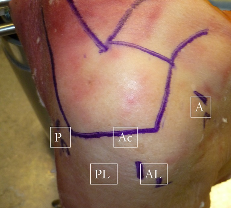
Fig. 1.
External view of the shoulder showing the standard portals used in repairing anterosuperior rotator cuff tears. For the working portion of the case, the arthroscope is placed in the posterolateral portal (PL). When working anteriorly either on the subscapularis or the LHBT, a penetrating device such as a “bird’s beak” can be placed through the anterior portal (A) while a grasper is placed through the anterolateral portal (AL) to aid in manipulating the tissue and feeding the sutures into the penetrator. As the repair progresses superiorly into the supraspinatus, anchors can be placed through a percutaneous portal on the lateral edge of the acromion (Ac). The scope is left in the PL portal, and an automatic suturing device is placed through the AL portal while sutures are retrieved through the A portal. The repair proceeds from anteroinferior to posterosuperior. The sutures are then tied from posterior to anterior.

