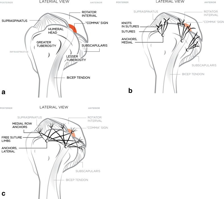
Fig. 6.
Schematic representation of the technique as seen from a lateral view. The bridging “comma sign” tissue between the supraspinatus and subscapularis is maintained (a). The supraspinatus, subscapularis, and bridging “comma sign” tissue are then brought over as a single cuff of tissue to a medial row of suture anchors with one anchor in the bicipital groove allowing for concomitant biceps tenodesis (b). The free suture limbs are then placed through knotless anchors forming a lateral row (c).

