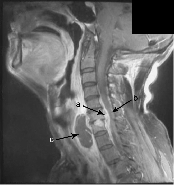
Fig. 2.
Cervical sagittal T1-weighted gadolinium-enhanced MR image performed on admission shows (a) an extensive spinal epidural abscess (SEA) extending from C4 to T1 anterior to the spinal cord; (b) spinal cord myelopathy can be noted from C5 to C7 due to anterior compression by the lesion (black arrow); (c) also, it shows the infection of C6–C7 intervertebral disc linked with the epidural lesion and a prevertebral abscess with hypointense content and homogeneous enhancement of wall and septa.

