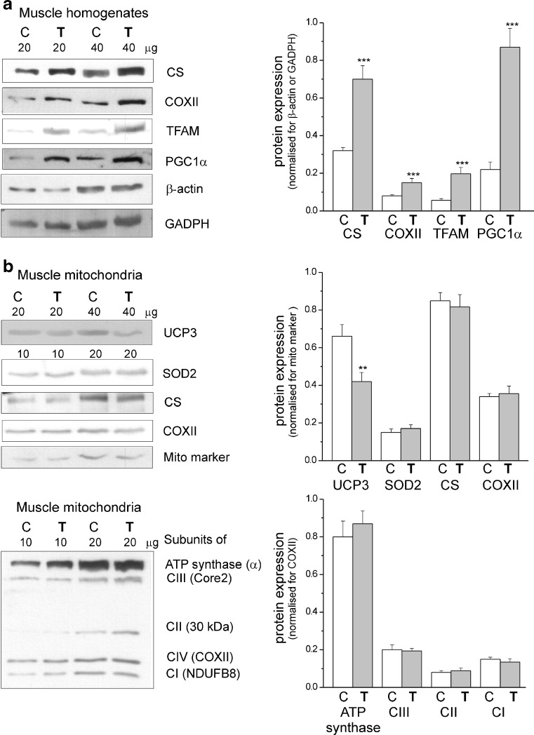
Fig. 2.
Determination of protein levels in skeletal muscle homogenates (a) and mitochondria (b) from control (C) and trained (T) rats. a Representative Western blots and analyses of the protein expression of CS, COXII, TFAM, PGC1α, β-actin, and GADPH. b Representative Western blots and analyses of the protein expression of UCP3, SOD2, CS, COXII, mitochondrial marker (mito marker), and particular subunits of ATP synthase, complex III (CIII), complex II (CII), and complex I (CI). Expression levels normalized for β actin (a), mito marker (b, upper panel), or COXII (b, lower panel) protein abundance are shown (a, b, right panels). The data (±SD, n = 12) are from six independent homogenate or mitochondrial preparations (duplicate assays for each experiment). Asterisks, comparison vs. value obtained for control rats

