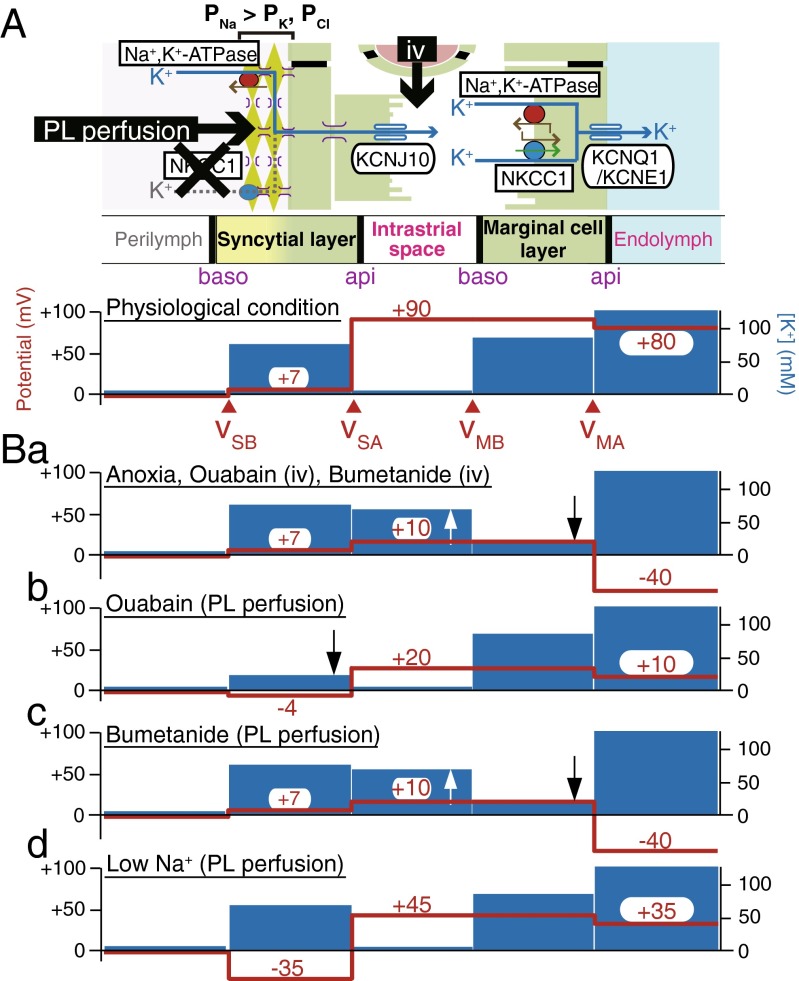
Fig. 3.
Summary profiles of the electrochemical properties of the lateral wall. In A, the upper panel illustrates the cellular compartments of the lateral wall and the channels and transporters contributing to unidirectional K+ transport and EP; the lower panel depicts the potential (red) and [K+] (blue) in intracellular and extracellular spaces under physiological conditions. Intravenous application (iv) of artificial solutions containing chemical compounds or modified ion compositions may primarily affect channels and transporters on the apical surface (api) of the syncytial layer and on the basolateral surface (baso) of the marginal cell layer, whereas perilymphatic (PL) perfusion is likely to strongly influence proteins on the basolateral surface of the syncytial layer. v SB, v SA, v MB, and v MA indicate the membrane potentials across the basolateral and apical surfaces of the syncytial layer and across the basolateral and apical surfaces of the marginal cell layer, respectively. The panels in B display the potential and [K+] properties observed during anoxia, vascular perfusion of ouabain or bumetanide (a), perilymphatic perfusion of ouabain (b), bumetanide (c), and a low Na+ solution (d). Observation of b, c, and d implies that the Na+,K+,2Cl−-cotransporter type 1 (NKCC1) present on the basolateral surface of the syncytial layer is unlikely to transport K+ and that this membrane domain is more permeable to Na+ than K+ and Cl− (P Na > P K, P Cl), as shown in the upper panel of A [115, 116]. The upward and downward arrows indicate the increase and decrease of [K+]. Reproduced and modified from [1, 74, 115, 116]

