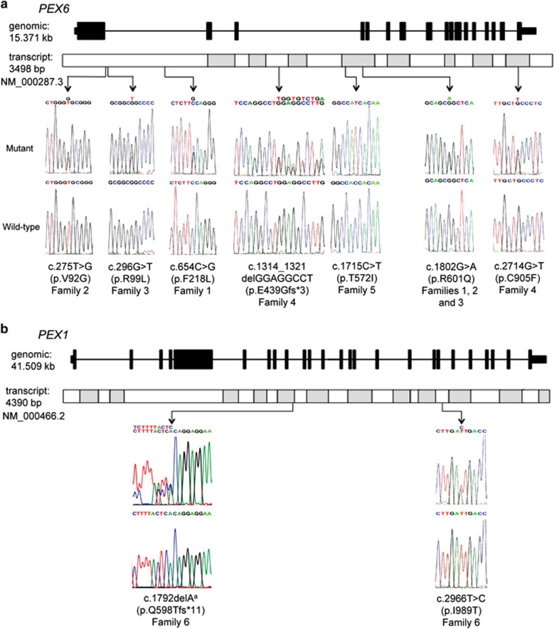
Figure 2.
Sanger sequencing and genomic locations of the mutations identified in this study. (a) A schematic diagram of PEX6 genomic structure and transcript shows the location and sequence traces of seven mutations identified in this study. (b) A schematic diagram of PEX1 genomic structure and transcript shows the location and sequence traces of two mutations identified in this study. aThe reverse sequence trace is shown for the PEX1 c.1792delA variant.

