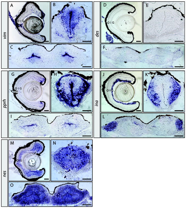
Figure 5. Expression patterns of vim, des, prph, ina, and nes in the pre-metamorphic tadpole nervous system.
In situ hybridization on retinal, brain and spinal cord sections for vim (A–C), des (D–F), prph (G–I), ina (J–L), and nes (M–O). Sections were obtained from pre-metamorphic stage 50 tadpoles. Location of the lens (L), outer (O), inner (I) and ganglion (G) cell layers are indicated. Scale bars, 100 μm in A, D, G, J and M; all others are 50 μm.

