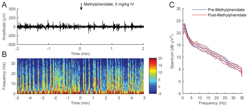
Figure 5. Methylphenidate does not induce significant electroencephalogram changes during continuous inhalation of 2.0% isoflurane.
(A) A representative electroencephalogram recorded from a rat inhaling 2.0% isoflurane, with time=0 indicating the start of methylphenidate (5 mg/kg IV) injection. The electroencephalogram remained in a burst suppression pattern after methylphenidate was administered. (B) A spectrogram computed from the same animal shows that methylphenidate did not induce significant changes in spectral power. (C) Power spectral densities computed from burst periods 2-minutes pre- and post-methylphenidate, and their 95% confidence intervals (with Bonferroni correction), were constructed around the mean spectra across all rats (n = 6). Methylphenidate did not induce significant changes in the power spectrum.

