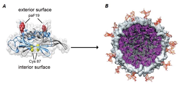
Figure 3.
Basic structure of MS2 nanocarriers: A) the external surface of the MS2 coat displays the modified aminoacid p-aminophenyllalamine(paF19) and the protein on the internal phage surface displays cysteine residues (Cys 87). B) After removing the phage genome, the Cys 87 is modified with a maleimide-conjugated porphyrin, and a Jurkat leukemia T cell-specific DNA aptamer is attached to the paF19. After the carrier enters into the targeted cell and is illuminated, reactive oxygen species are produced and the cancerous cells are disrupted. (Reprinted from ref.[72] Copyright (2016) American Chemical Society).

