
Figure 2.
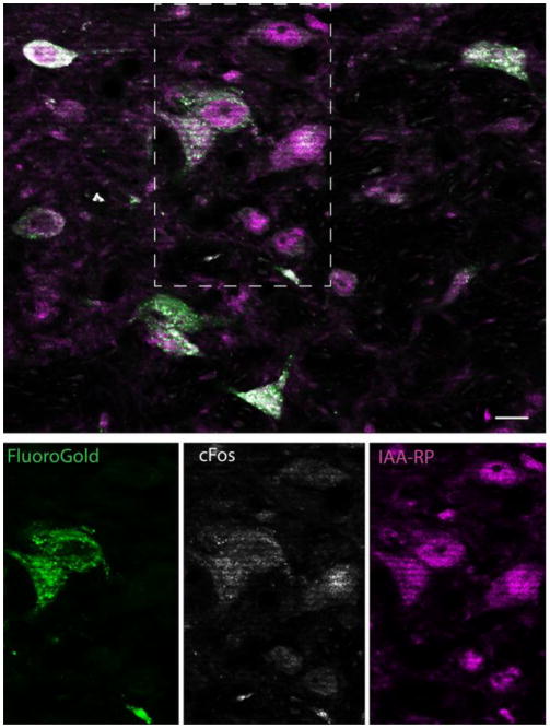
Overview of vestibular neurons with direct projections to the ventrolateral medulla (FluoroGold-immunopositive), which were activated by sGVS (cFos-positive) and co-localized IAARP. Top panel shows a cluster of single, double and triple-labeled neurons in ipsilateral SpVN from a rat with a FluoroGold injection in RVLM. The individual channel images of the area delineated by the dashed rectangle are shown in the panels below. These single channel images illustrate co-localization of cFos and FG in two neurons, the activation of one additional neuron that was not retrogradely filled, and the presence of IAARP in all three of those cells, as well as others in the field. Scale bar is 10 μm.