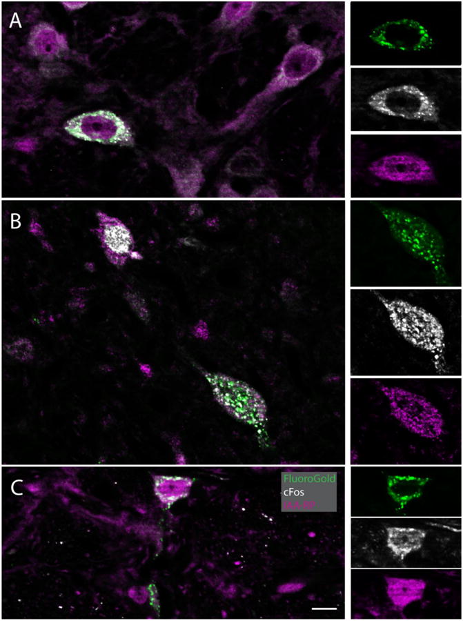
Figure 3.

IAARP is present in the cell bodies of sGVS-activated vestibular neurons with direct projections to the ventrolateral medulla. Each large panel (A-C) illustrates one triple-immunofluorescent vestibular neuron, as well as several double and single labeled cells. The side panels are single channel images of the triple-labeled neurons in the larger panels to the left. For each set of side panels, FluoroGold label is shown at the top, cFos in the center, and IAARP at the bottom. A: A multipolar neuron in SpVN with projections to contralateral CVLM. B: A fusiform neuron in MVN that projects to ipsilateral RVLM. The smaller neuron in the upper left quadrant of the panel is cFos and IAARP-labeled, but did not contain retrograde tracer. C: A small spherical neuron in SpVN with projections to ipsilateral RVLM. Scale bar is in C is 10 μm for all large panels. The single channel images are slightly enlarged.
