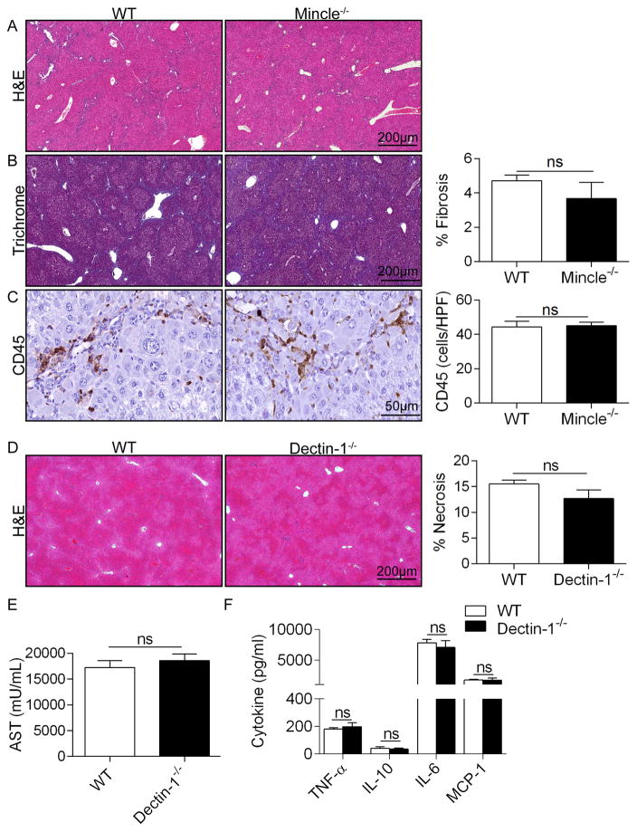
Figure 4. Mincle deletion does not mitigate TAA-induced liver fibrosis and Dectin-1 deletion does not protect against ConA hepatitis.
(a–c) WT and Mincle−/− mice (n=5/group) were serially treated with TAA for 12 weeks. Livers were examined by (a) H&E and (b) trichrome staining and the percent fibrotic area was calculated. (c) CD45+ pan-leukocyte infiltrate were determined by immunohistochemistry. (d–f) WT and Dectin-1−/− mice were treated with Con-A (20μg/g). (d) Livers were harvested at 12h and examined by H&E staining. Representative H&E-stained sections are shown and the fraction of non-viable liver was calculated. (e) Serum levels of AST, (f) TNF-α, IL-10, IL-6, and MCP-1 were calculated for each cohort (n=5 group; p=ns for all comparisons). In vivo experiemnts were repeated twice with similar results.

