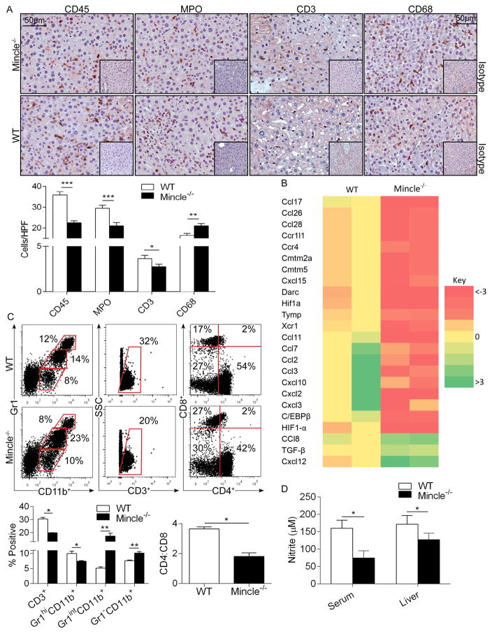
Figure 6. Mincle deletion modulates intra-hepatic inflammation and nitrite production in Con-A hepatitis.
(a–c) WT and Mincle−/− mice were treated with Con-A (20μg/g). (a) CD45+ pan-leukocyte, MPO+ neutrophil, CD3+ T cell, and CD68+ macrophage infiltrate were determined by immunohistochemistry at 12h. Results were quantified by examining 10 HPFs per slide (n=5/group). (b) Intra-hepatic inflammation was determined by mRNA levels of diverse inflammatory mediators in whole liver tisues. A heat map analysis showing fold change in Mincle−/− mRNA expression levels relative to WT controls is shown (n=2/group). (c) Cellular ractions of myeloid and T cell subsets in livers of Con-A-treated WT and Mincle−/− mice were determined by flow cytometry. Representative dot plots and quantifications based on replicates, including the CD4:CD8 ratio, are shown. Flow cytometry experiments were reproduced more than 3 times using 3–5 mice per cohort. (d) The Griess assay was used to determine nitrite levels in the liver and serum of WT and Mincle−/− mice at 12h after Con-A administration. (n=5/group; *p<0.05, **p<0.01, ***p<0.001).

