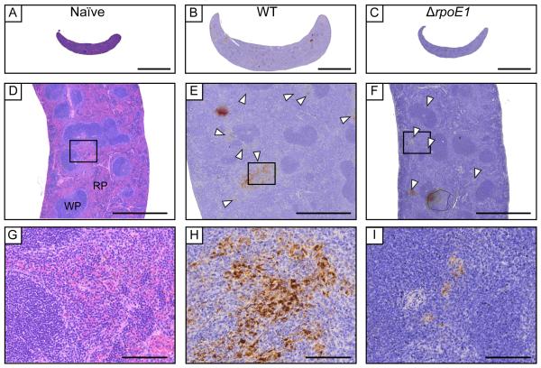
Figure 3. Spleen histology of BALB/c mice infected with wild-type and ΔrpoE1 B. abortus strains.
At 9 weeks post-infection spleens were harvested, fixed, and slides were prepared. H&E staining was performed on all samples. Uninfected (A, D, G), WT-infected (B, E, H), or ΔrpoE1-infected (C,F,I) mouse spleen pictures were taken at 10X (A-C), 50X (D-F) or 400X magnification (G-I). Brucella antigen was visualized by immunohistochemistry with an anti-Brucella antibody (D-I). White arrowheads point to areas of extensive Brucella antigen staining. Scale bars are 5 mm (A), 1 mm (B), and 100 μm (C). WP = white pulp, RP = red pulp.

