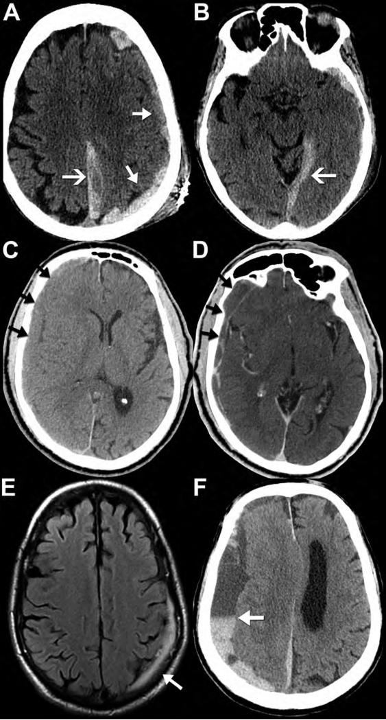
Figure 4. CT and MR appearance of subdural hematomas.
Noncontrast CT (A,B) performed on a 90-year-old female after fall with left parietal scalp laceration reveals hyperdense blood along the left convexity (A, closed arrows), left posterior falx cerebri (A, open arrow), and tentorium cerebelli (B, open arrow). Note how the subdural hematomas do not cross the dural sinuses to the other side of the falx or tentorium. Noncontrast CT performed on an 82-year-old man after falling shows a right convexity subacute subdural hematoma that is isodense to the adjacent cortex (C, arrows). Post-contrast imaging is generally not required for evaluation of a subdural hematoma; however, it was obtained in this case. The post-contrast CT (D, arrows) images demonstrate peripheral enhancement of the collection without evidence of active extravasation, thus reinforcing a subacute injury. Axial FLAIR MR image (E) from a 69-year-old man after head trauma exemplifies the high contrast difference on MR between the FLAIR hyperintense subdural hematoma (arrow) and the adjacent hypointense calvarium. Axial noncontrast CT (F) from a different patient, a 73-year-old man with left-sided weakness, shows the appearance of a mixed-density, “acute-on-chronic” subdural hematoma along the right convexity and falx cerebri. Note the dependent layering of the acute, denser blood products within the chronic, hypodense collection; this is often referred to as the “hematocrit sign” (arrow).

