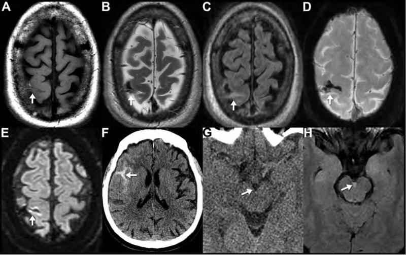
Figure 5. CT and MR appearance of subarachnoid hemorrhage.
MRI (A-E) performed on a 58-year-old man who presented 3 days after a fall with altered mental status. Subacute subarachnoid hemorrhage appears hyperintense to brain on T1WI (A) and hypointense on T2WI (B). Subarachnoid hemorrhage does not suppress like normal CSF on FLAIR imaging (C) and it appears markedly hypointense on SWI (D). The linear area of reduced diffusion in the cortex adjacent to the subarachnoid hemorrhage likely represents adjacent cerebral contusion (E). This is a good example showing the appearance of early subacute subarachnoid blood products within the central sulcus on multiple pulse sequences (arrows). Noncontrast CT (F) from a 70-year-old male after syncope and fall with head injury shows the classic appearance of acute subarachnoid hemorrhage in the right sylvian fissure (arrow). Noncontrast CT (G) and FLAIR (H) images obtained the same day in a 33-year-old female after a motor vehicle crash. The trace interpeduncular subarachnoid hemorrhage (arrow) is invisible on CT (G), but is readily apparent on MR (H), highlighting the greater sensitivity of MR to blood products.

