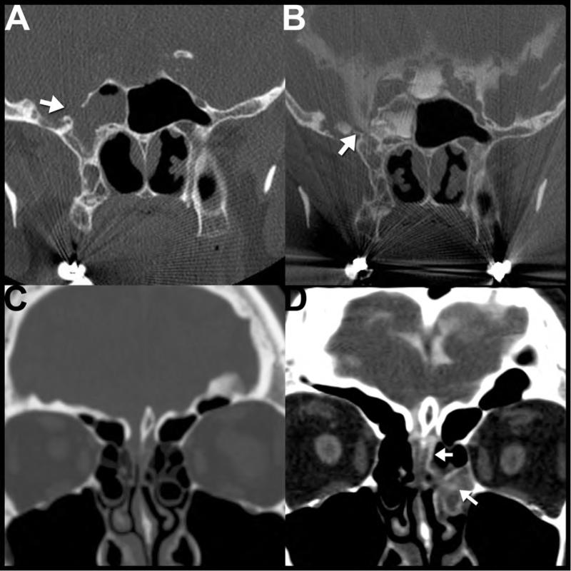
Figure 6. Imaging evaluation of patients with CSF leaks.
Coronal reformatted images from a noncontrast CT (A,C) and CT cisternograms (B,D) performed in 2 different patients with posttraumatic CSF leaks. The first patient (A,B) is a 55-year-old female with a history of remote trauma and meningitis. Note the opacified right sphenoid sinus with a large bony defect between the lateral wall of the right sphenoid sinus and the middle cranial fossa (A, arrow). CT cisternography was performed (B), confirming abnormal leakage of CSF contrast from the middle cranial fossa into the sphenoid sinus. The second patient (C,D) is a 33-year-old female with CSF rhinorrhea (confirmed with positive B2-Transferrin test) after facial trauma. Noncontrast CT (C) reveals subtle unilateral opacification of the left olfactory recess concerning for a possible cephalocele, but it did not show a definitive bony abnormality in that region. The patient returned two weeks later and a CT cisternogram was performed (D) revealing abnormal passage of CSF contrast (arrows) through the left cribiform plate through the left olfactory recess and into the nasal cavity.

