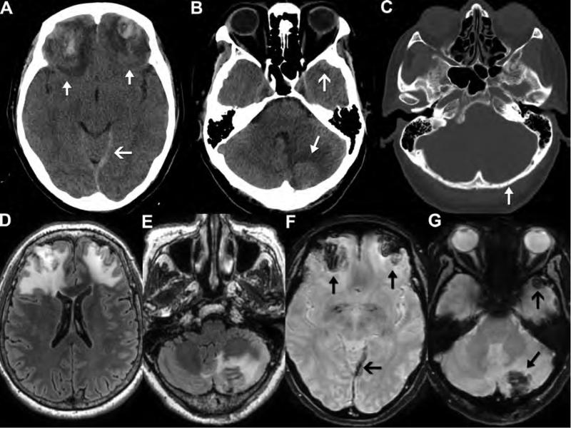
Figure 7. CT and MR appearance of cerebral and cerebellar contusion.
Noncontrast CT (A-C) and FLAIR and susceptibility-weighted MR (D-G) images obtained on a 59-year-old woman after a fall that occurred 5 days earlier. CT images show a nondisplaced left occipital fracture (C, arrow) with an underlying hemorrhagic contusion in the left cerebellar hemisphere (B, closed arrow) compatible with coup injury. There are also large, bifrontal, hemorrhagic contusions (A, closed arrows) and a small left anterior temporal hemorrhagic contusion (B, open arrow), compatible with contrecoup injuries. These lesions have a predictable appearance on MR with low signal on SWI (F, closed arrows; G, arrows) and a large area of surrounding edema on FLAIR images (D,E). Also note the increased prominence of the left tentorial subdural hematoma (open arrow) on MR SWI (F) compared to CT (A).

