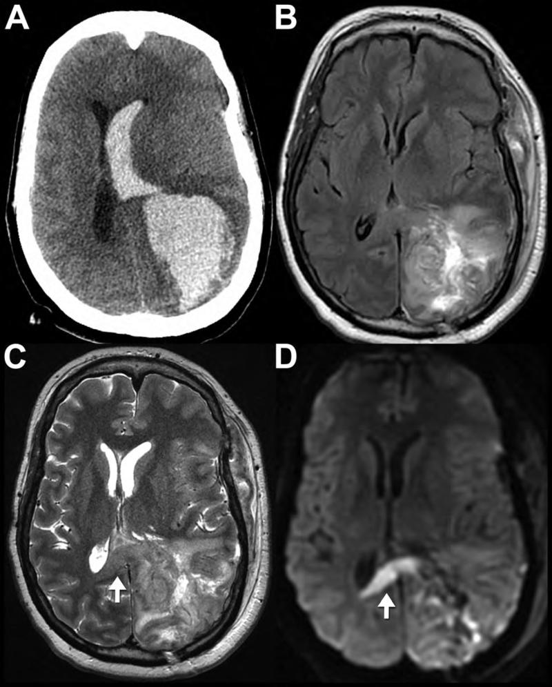
Figure 8. CT and MR appearance of intracerebral hematoma.
Noncontrast CT (A) of 61-year-old male after head trauma reveals a large, hyperdense, acute left parietal hematoma with surrounding edema that has decompressed into the adjacent left lateral ventricle (intraventricular hemorrhage). MRI performed 11 days later (B-D) shows decreased edema with residual T2 hyperintense blood products (likely corresponding to “late subacute” extracellular methemoglobin). Increased T2 signal intensity (C) and reduced diffusion (D) in the splenium of the corpus callosum (arrows) is likely a result of Wallerian degeneration of the axons leading away from the left parietal injury, and not traumatic axonal injury of the splenium.

