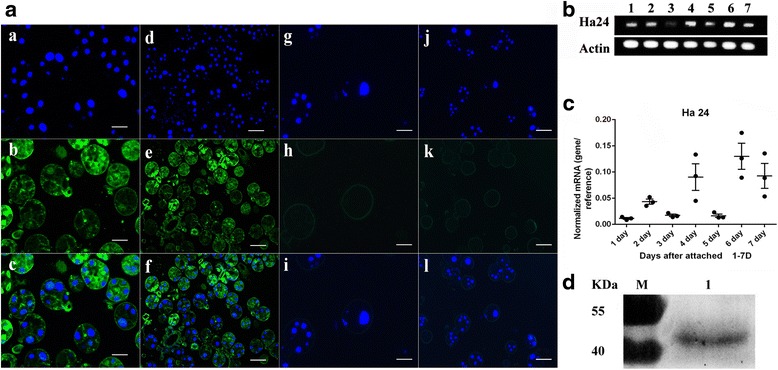
Fig. 5.

Identification of native Ha24. a Immunofluorescence analysis (IFA) of Ha24 in feeding tick salivary glands. Tick salivary glands were stained with mouse anti-Ha24 PcAb (a-f) and wild mouse PcAb (control, g-l), and DNA were stained with DAPI. a, d, g, j were observed in blue fluorescent (DAPI); b, e, h, k were observed in green fluorescent (FITC); a and d, b and e; g and h; and j and k were merged (shown in c, f, i and l, respectively). Scale-bars: a-c, g-i, 100 μm; d-f, j-l, 50 μm. b RT-PCR analysis of Ha24 in tick salivary glands (1–7 days after attached). 1 % agarose gel showing the Q-PCR products from feeding female tick salivary glands. Lanes 1–7: cDNA of tick salivary glands at different feeding periods with Ha24 specific primers; Lanes 1–7 below: cDNA of tick salivary glands at different feeding periods with tick actin primers. c Q-PCR analysis of Ha24 in tick salivary (1–7 days after attachment). The relative expression of Ha24 mRNA vs that of actin mRNA was investigated by quantitative reverse transcription polymerase chain reaction in different tick feeding stages. d Identification of native Ha24 by western blot. Lane M, pre-stained protein ladder; Lane 1, anti-Ha24 PcAb from feeding female tick salivary glands
