Fig. 1.

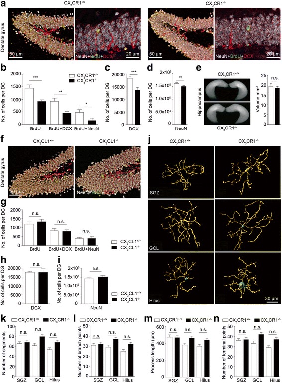
DCX+ cell numbers in the subgranular zone (SGZ) of the dentate gyrus (DG) are regulated by the fractalkine receptor, CX3CR1, but not by its ligand, fractalkine, under non-stimulated conditions. a Representative immunofluorescent images of coronal DG sections from cx3cr1 +/+ (left) and cx3cr1 −/− (right) mice stained for the neuronal marker NeuN (white), the proliferation marker bromdesoxyuridin (BrdU, green) and DCX (red). b Quantification of the number of BrdU (p < 0.0001, two-tailed Student’s t-test, cx3cr1 +/+ (n = 14) vs. cx3cr1 −/− (n = 13)) BrdU + DCX (p = 0.0029, two-tailed Student’s t-test, cx3cr1 +/+ (n = 13) vs. cx3cr1 −/− (n = 13)) and BrdU + NeuN (p < 0.0444, two-tailed Student’s t-test, cx3cr1 +/+ (n = 10) vs. cx3cr1 −/− (n = 10)) positive cells in DG revealed lower cell numbers in cx3cr1 −/− mice than in cx3cr1 +/+ mice. Similarly, cx3cr1 −/− mice had reduced total numbers of DCX+ (p < 0.0001, two-tailed Student’s t-test, cx3cr1 +/+ (n = 14) vs. cx3cr1 −/− (n = 13)) (c) and NeuN+ cells when compared to cx3cr1 +/+ controls (p = 0.0048, two-tailed Student’s t-test, cx3cr1 +/+ (n = 9) vs. cx3cr1 −/− (n = 9)) (d) even though the overall hippocampal volume was not different in both mouse lines (p = 0.6362, two-tailed Student’s t-test, cx3cr1 +/+ (n = 4) vs. cx3cr1 −/− (n = 4)) (e). f Representative immunofluorescent images of coronal DG sections from cx3cl1 +/+ (left) and cx3cl1 −/− (right) mice stained with antibodies against NeuN (white), BrdU (green) and DCX (red). g In the DG of both mouse strains cell numbers for BrdU, BrdU + DCX and BrdU + NeuN positive cells where not statistically different. There was further no difference in the cell numbers quantified for DCX+ (h) and NeuN+ cells (i) (p > 0.05, two-tailed Student’s t-test, cx3cl1 +/+ (n = 9) vs. cx3cl1 −/− (n = 9)). j 3D-reconstruction of ionized calcium-binding adapter molecule 1 (Iba-1)-labeled microglia from cx3cr1 +/+ (left) and cx3cr1 −/− (right) mice localized in the SGZ (top row), granular cell layer (GCL, middle row) and hilus (bottom row) of the DG. There were no significant morphometric differences in microglia of both mouse lines after quantifying k number of segments, l number of branch points, m process length and n number of terminal points (p > 0.05, two-tailed Student’s t-test, cx3cr1 +/+ (n = 25–31 cells from 4 animals) vs. cx3cr1 −/− (n = 31–37 cells from 4 animals)). One representative experiment of two is shown. Bars represent mean ± SEM; n.s., not significant. ***p < 0.001; **p < 0.01; *p < 0.05
