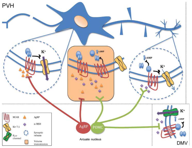
Figure 4.
A new model of MC4R microcircuitry. The Yin–Yang model of α-MSH and AgRP action (Fig. 1) suggested competitive binding of these peptides to individual MC4R sites (orange box), and anatomical data suggest in regions where these peptides undergo volume release, that competition for binding to the MC4R may occur. New subcellular anatomical data suggest that in the PVH, AgRP synaptic contacts predominate at cell bodies, while POMC synaptic contacts predominate at distal dendrites. Along with the fact that AgRP immunoreactive fibers are only observed in a subset of MC4R expressing nuclei containing POMC-immunoreactive fibers, α-MSH may this often act independently of AgRP (right circle). At these sites, α-MSH may be expected to signal through both cAMP, and Kir7.1. The ability of AgRP to act independently of α-MSH as a potent hyperpolarizing agonist, via regulation of Kir7.1, suggests the likely existence of independent AgRP sites of action (left circle). Another MC4R signaling pathway, involving cAMP/PKA-dependent activation of KATP channels and α-MSH-induced hyperpolarization, has been demonstrated in MC4R neurons in the dorsal motor nucleus of the vagus in the brainstem (bottom right). Thus, α-MSH and AgRP utilize a diversity of signaling modalities to regulate feeding and energy homeostasis through the MC4R. Modified, with permission, from Ghamari-Langroudi M, Digby GJ, Sebag JA, Millhauser GL, Palomino R, Matthews R, Gillyard T, Panaro BL, Tough IR, Cox HM, et al. (2015) G-protein-independent coupling of MC4R to Kir7.1 in hypothalamic neurons. Nature 520 94–98.

